Endoscopic mitral valve surgery

Mitral valve
The mitral valve is located between the left atrium and the left ventricle. Oxygen-enriched blood from the lungs passes through it into the left ventricle. Its main function is to control the blood flow during the cardiac cycle by closing during the contraction of the heart and opening during the relaxation phase. This ensures that the blood only flows in one direction during the pumping process - from the atria to the ventricles. If the mitral valve suffers from a leak (mitral valve insufficiency), the valve can no longer ensure a stable blood flow and the blood returns to the lungs, leading severe symptoms such as shortness of breath, palpitations and reduced exercise capacity. If the mitral valve is narrowed and can no longer open sufficiently (mitral valve stenosis), the blood flow from the left atrium into the left ventricle is obstructed. This leads to shortness of breath, rhythm disturbance with reduced performance.
Mitral valve reconstruction
If the mitral valve is diseased and can no longer adequately fulfill its function, such as in the case of mitral valve insufficiency, surgical mitral valve reconstruction is necessary.
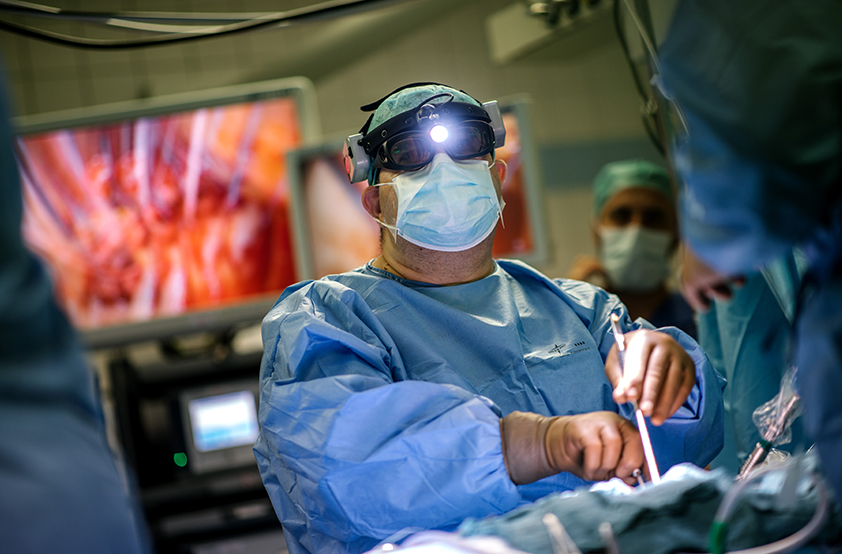
Endoscopic mitral valve repair is a minimally invasive procedure for treating diseases of the mitral valve. This technique allows the surgeon to reconstruct the mitral valve without the need for a large chest incision. A 3D camera allows the surgeon to visualize the entire surgical field on a screen through 3D glasses. The valve can be repaired using special surgical instruments.
The repair of the mitral valve can involve various techniques, such as suturing or tightening tissues, removing excess tissue or placing support elements to stabilize the valve and restore its function for many years into the future.
In endoscopic mitral valve reconstruction, a four-centimeter incision is performed between the right-sided ribs, at the level of the fourth intercostal space or as a crescent-shaped incision around the nipple. The heart-lung machine, which takes over the oxygen supply to the other organs during the operation and stopping of the heart, is connected via the femoral vessels or the subclavian artery.
Endoscopic mitral valve surgery is a promising surgical technique and can help to improve the recovery and quality of life of patients with mitral valve disease for a longer period of time. In addition, the patient's natural mitral valve is preserved, resulting in improved long-term cardiac function.
Endoscopic mitral valve replacement
Endoscopic mitral valve replacement is a minimally invasive technique in which the diseased mitral valve can be replaced with a biological (bovine/pig) or mechanical prosthesis via a minimally invasive approach. In an endoscopic mitral valve replacement, the surgeon inserts small instruments and a 3D camera through small incisions in the patient's chest wall. Access is gained via a four-centimeter incision between the right-sided ribs at the level of the fourth intercostal space. The special small surgical instruments enable the surgeon to replace the mitral valve, and the 3D camera gives the surgeon a highly defined view of the entire surgical field with the aid of 3D glasses. The heart-lung machine, which takes over the oxygen supply to the other organs during the operation and stopping of the heart, is mainly connected via the femoral vessels or the subclavian artery.
This method offers several advantages over traditional open heart surgery, including shorter recovery times, less post-operative pain, and a quicker return to normal activities.
